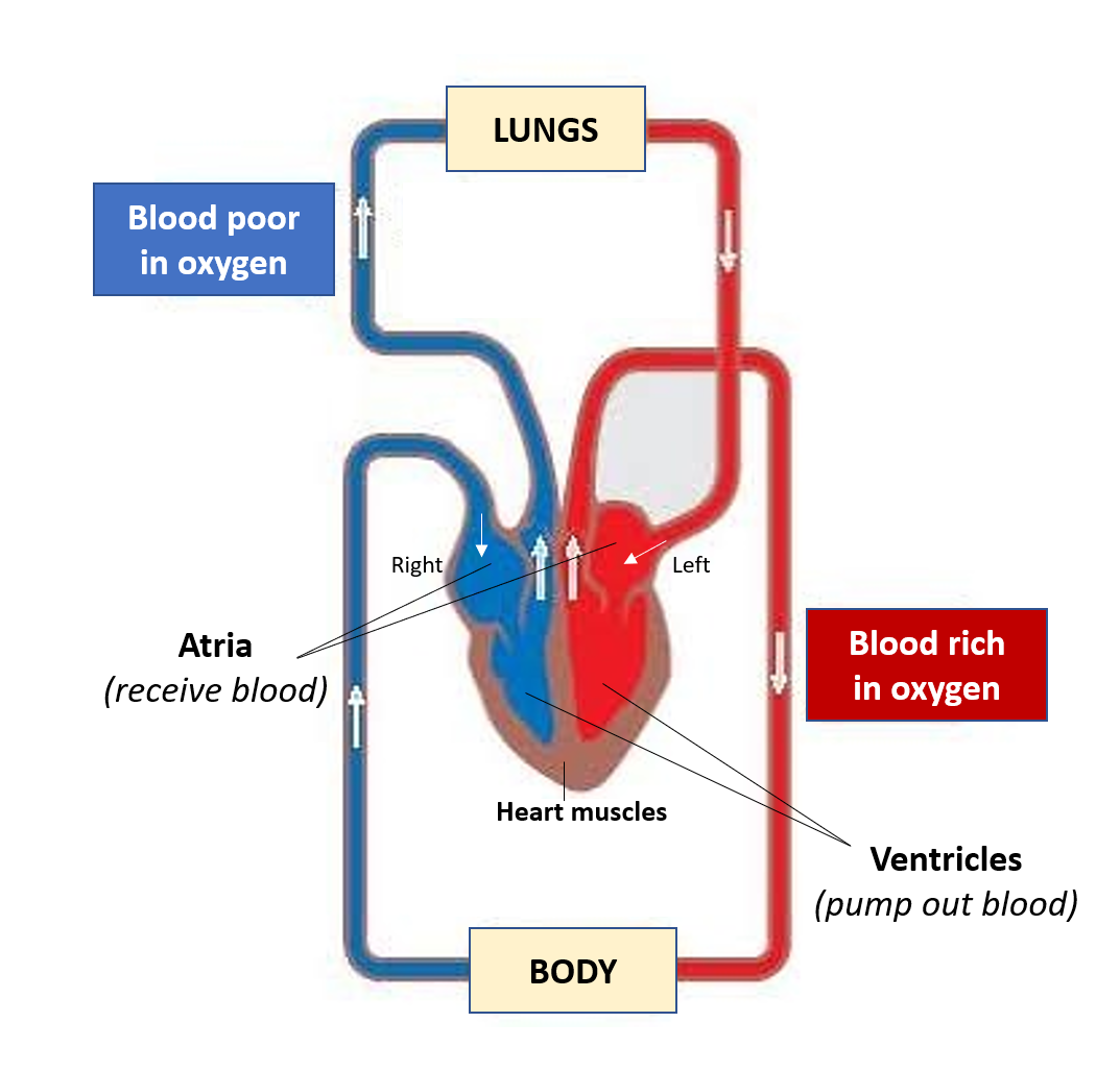
Our heart works tirelessly and quietly round the clock, so evidently if it gets ill in any way, the health impact can be serious and significant. Even though the heart chambers (atria and ventricles) are filled with blood, the cardiac muscles (myocardium) require blood supply and oxygen (provided by coronary arteries), to effectively contract and pump the blood to the entire body.
A lot of terms associated with heart (cardiac) conditions are used in common parlance but it is useful to be aware of what these terms really mean.
Atherosclerosis/Arteriosclerosis
This refers to the buildup or formation of plaques in the blood vessels called arteries which carry oxygen-rich blood to all the body organs. These plaques (also called atheroma) are made up of Fats, Cholesterol, Calcium, and some blood cells mainly the Platelets. Repeated ‘micro-injuries’ to the artery wall and increased levels of fats (cholesterol and triglycerides) transported in blood predispose to plaque formation. The plaques can cause partial or complete blockage of the arteries.
CARDIOVASCULAR DISEASE (CVD)
CVD, also known as Atherosclerotic Cardiovascular Disease (ASCVD), refers to conditions caused by the blockage of blood vessels (arteries) due to atherosclerosis. People at risk of developing atherosclerosis and CVD include patients with high BP (hypertension), diabetes, obesity, sedentary lifestyle, smoking, high carbohydrate/sugar and/or saturated/trans-fat intake in the diet or those who show high lipids (fats like cholesterol/triglycerides) levels in the blood. Risk increases with age and if there is a similar history in the family.
When blood flow (and therefore oxygen supply) is reduced to the heart, it is called Ischemic Heart Disease (IHD, Ischemia means lack of oxygen) or Coronary Artery Disease (CAD- blockages in the coronary arteries which supply the heart). If more than 50% of any coronary artery is blocked, it is called ‘obstructive’ CAD.
Angina
The lack of adequate oxygen supply to any part of the heart leads to a lack of functioning of that part, and heart pain called ‘angina’.
Stable Angina
In people with obstructive CAD, the heart may continue to get enough blood and oxygen at rest or during normal activities. But during exertion, stress, exercise, or illness, there is a need for more powerful pumping and increased oxygen requirement of the heart. At this time there may be inadequacy in blood and oxygen supply to parts of the heart muscles (which do the pumping action), due to the artery blockages. This results in heart pain called stable angina. Stable angina is relieved with rest and giving medicines (nitrate drugs) to further dilate these partially blocked arteries and reduce or cope with the oxygen demand of the heart.
Unstable Angina
Sometimes a plaque causing partial obstruction of the artery ruptures periodically and the resulting blood clot may majorly obstruct the blood supply to a portion of the heart. Such plaques are called unstable plaques. Since this can happen at rest or even on mild exertion, it is unpredictable and called unstable angina. This kind of angina is not relieved by rest, and nitrate dilators may also not relieve the pain completely or adequately.
Myocardial Infarction
When an almost complete, or complete blockage is present in any coronary artery, it can cause the stoppage of blood flow and oxygen supply to that part of the heart. This is called Myocardial Infarction (MI) or Heart Attack. MI also causes severe heart pain, and again this pain is not relieved either by rest (can even occur at rest) or by dilating medicines. (MI is further classified into ST-segment elevation- STEMI and Non-ST-segment elevation- NSTEMI, based on changes seen in ECG).
Unstable angina and MI are together called Acute Coronary Syndrome (ACS). The pain usually lasts more than 10 minutes, compared to the usual 2-5-minute duration seen with stable angina. The pain is like a squeezing or tightening sensation in the chest which may radiate to the jaw, neck, arm, shoulder, and back. Often symptoms of anxiety-like breathlessness, lightheadedness, sweating, and nausea may be associated. Sometimes the pain may not be so typical and get dismissed as indigestion or gas, so it is best to do an ECG in case of the slightest suspicion.
It may be hard to clinically differentiate unstable angina and MI. ECG changes and rising cardiac enzymes in the blood (markers of MI) can help establish the diagnosis.
Angina or MI can be the first manifestation of CAD, therefore it is important to have your BP, blood lipids (triglycerides and cholesterol), and sugar checked periodically if you have any of the risk factors mentioned above, or are more than 45 years (male) or 55 years (female) years of age. You may be advised further tests like ECG and 2D-echocardiography to assess cardiac function, and angiography to diagnose the extent of blockage of arteries.
Treatment of high BP and diabetes, diet modification and weight management, regular exercise, and cessation of smoking would be the steps advised. In addition, to reduce plaque formation, medicines to lower cholesterol/triglycerides, and prevent clots in the arteries (antiplatelet drugs like aspirin, clopidogrel) are also prescribed
Treatment of an acute coronary episode requires immediate as well as long term blood thinning/anti-platelet medicines (heparin group, aspirin, clopidogrel) with timely hospitalization (preferably in a 1-hour window). Interventions on the blocked vessels include angioplasty with stenting or bypass grafting depending on the type and extent of blockages. Risk factor management and dietary modifications would always be needed lifelong.
Blockages of other vessels can lead to diseases in other organs like the brain (stroke), limbs (peripheral vascular disease – PVD), and kidney disease (Atherosclerotic Reno-Vascular disease ARVD).
HEART FAILURE
Heart failure (HF), also called Congestive Cardiac Failure (CHF) occurs when the heart is unable to pump adequate blood into the arteries, and the backlog of blood in the heart leads to congestion and backpressure into the lungs and the veins.
Ejection fraction (EF), measured by echocardiography, is the percentage of how much blood the left ventricle pumps out with each contraction. Based on this, HF is of the following types:
- Heart failure with reduced ejection fraction (HFrEF) or systolic heart failure: EF is 40% or less, signifying deficient pumping action possibly due to weak or damaged cardiac muscles.
- Heart failure with preserved ejection fraction (HFpEF) or diastolic heart failure: EF is 50% or more, signifying that even though pumping is effective, there is a deficient filling of the ventricle, possible due to muscle stiffness resulting from disease, damage or scarring.
- EF of 41-49% is considered borderline.
Causes of HF include an episode of a massive MI where large parts of the cardiac muscles are damaged due to oxygen deprivation. It can also occur due to prolonged hypertension as well as CAD, as there is an increased load on the heart, pumping against higher pressure and resistance. Other causes include viral and degenerative diseases of the muscles (myocarditis, cardiomyopathy), abnormalities/diseases of the cardiac valves, or rhythm disturbances.
Symptoms of HF are:
- breathlessness (especially on lying down) or on exertion
- swelling of legs/ankles/feet/abdomen/face-eyelids
- weakness/tiredness
- palpitation
- lack of appetite
- increased need to pass urine (especially at night)
Medical management includes medicines to decrease the cardiac load by improving BP control and heart function (ACE inhibitors/ARBs), dilating the arteries and the veins (vasodilators like CCB group of drugs), decreasing heart rate (Beta-blockers), and eliminating excess fluid (diuretics). These are also drugs used to manage high BP. New drugs called angiotensin receptor neprilysin inhibitors (ARNI: Sacubitril-Valsartan) are also available now for heart failure not responding well to other medicines. Oxygen therapy may be needed in people who have significant congestion of blood in the lungs, due to reduced pumping of the left side of the heart.
CARDIAC ARREST
It is also called sudden cardiac death. It implies that the heart has stopped pumping blood.
Cardiac arrest is always the final cause of death in patients with heart diseases like heart attack, heart failure or electric rhythm disturbances. It is also the ultimate cause of death in drowning, electrocution, poisoning/drug overdose, as well as massive – injuries, blood loss, and infections (causing shock).
Often in conversations, the words heart attack, cardiac arrest, and heart failure are used interchangeably, but the awareness that they refer to three different clinical situations is important. Also, a heart attack can be the cause of heart failure which eventually causes a cardiac arrest, or a massive heart attack can be the direct cause of a cardiac arrest, which is the final cause of death.
One should perform a CPR (Cardio-Pulmonary Resuscitation) which should be ideally initiated within 2-5 minutes in a person with cardiac arrest. This can help restart the pumping action of the heart and revive the patient while emergency services are on their way. After 6-8 minutes, permanent brain damage is inevitable with patients often going into a coma. After 10 minutes, the chances of survival are extremely low.
Also read –
Diabetes: Understanding health risks and effective diet-lifestyle measures
For any query, additional information or to discuss any case, write to info@drvarsha.com and be assured of a response soon
References
2019 ACC/AHA Guideline on the Primary Prevention of Cardiovascular Disease