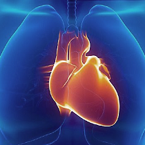
Carditis refers to the inflammation of one or more layers of the heart. It includes Pericarditis, Myocarditis, Endocarditis and Pancarditis.
The heart has 3 layers-
Pericardium: This is the outer protective layer. It is bi-layered with pericardial fluid in between that helps smooth movement during heartbeat/pumping by reducing friction, and has a cushioning effect.
Myocardium: This is the middle layer made up of muscles that cause the heart to contract and pump blood.
Endocardium: This is the inner layer lining the heart chambers and the cardiac valves with its associated structures like attachment chords and muscles (chordae tendineae and papillary muscles respectively).
PERICARDITIS
It refers to inflammation of the pericardium.
CAUSES
- Infections – Viral (most common cause), bacterial and rarely fungal
- After heart attack or heart surgery
- Injuries
- Other medical conditions: Kidney failure, AIDS, TB, cancer, radiation exposure
- Autoimmune diseases (Collagen Vascular Diseases): Systemic lupus erythematosus (SLE or Lupus), scleroderma, rheumatoid arthritis, polymyositis/ dermatomyositis, and Sjögren (dry eye/mouth) syndrome.
- Medicines: Like phenytoin (anti-epileptic), blood thinners (anticoagulants) warfarin and heparin, procainamide (for irregular heart beat -arrhythmia).
SYMPTOMS
Pain is the main symptom. It is sudden, stabbing and on the left side spreading sometimes to shoulder and neck. So, it can be confused with a heart attack. Pain gets worse on deep breathing or coughing, and on while lying down, while it may improve on sitting and leaning forward.
Other symptoms include cough, breathlessness on lying, palpitation, low grade fever, weakness/fatigue and swelling of feet or abdomen.
TYPES
Acute pericarditis begins suddenly and is of short duration (usually<3 weeks).
Recurrent pericarditis can occur after 4-6 weeks of an episode of acute pericarditis that has completely resolved.
Chronic pericarditis develops gradually and lasts longer than 12 weeks.
Some terms to describe the nature of pericarditis used are fibrinous (forming fibrous adhesions), exudative (with inflammatory fluid), and purulent (with pus).
COMPLICATIONS
Pericardial effusion (effusive pericarditis) refers to the build-up of fluid in the pericardium which starts putting pressure on the heart and affects its filling and pumping ability. This can result in heart failure, and cardiac tamponade (a medical emergency resulting in sudden blood pressure drop, breathlessness and dizziness that can even be fatal). The accumulated fluid has to be drained promptly by inserting a needle into the pericardium (pericardiocentesis or pericardial tap).
Constrictive pericarditis can be a sequel of chronic pericarditis due to thickening, scarring and stiffening of the pericardium leading to restriction in both filling and pumping of the heart. This can eventually cause heart failure, as well as heart rhythm problems (arrhythmia).
DIAGNOSIS AND TREATMENT
Pericarditis is diagnosed with clinical suspicion based on symptoms and examination, and tests like chest X-ray, ECG, echocardiography and a cardiac MRI.
Pericarditis often resolves on its own with rest or simple treatment. Anti-inflammatory medicines like NSAID group or a course of corticosteroids may be given, sometimes along with antibiotics if bacterial infection is suspected. In case of pericardial effusion, a pericardial tap to remove fluid with needle insertion is done.
In chronic cases, diuretics to reduce swelling may be needed, as well as medicines for heart failure and when needed drugs to manage arrhythmia. A stiff pericardium may need surgical removal (pericardiectomy).
MYOCARDITIS
Myocarditis is an inflammation of the middle layer of heart muscle (myocardium). This can affect the heart’s pumping ability and heartbeat (cause arrhythmia).
CAUSES
- Infections: Viral infection is the most common cause. Causative viruses include adenovirus causing common cold, flu (influenza virus), parvovirus, echovirus (causes gut infections), COVID, hepatitis, rubella, Epstein-Barr (causes infectious mononucleosis), herpes and HIV. Bacteria (staphylococcus, streptococcus, diphtheria and Lyme’s disease), parasites (toxoplasmosis, Trypanosoma – seen more in travelers) and fungi (candida, aspergillus, Histoplasma) are also more rare causes of myocarditis.
- Reactions to drugs/toxins: These include some drugs for cancer chemotherapy, anti-epileptics, antibiotics (penicillin/sulfonamides), cocaine and other drugs of abuse, radiation exposure, toxic gases like carbon monoxide, and sometimes to vaccines.
- Inflammations like vasculitis and autoimmune diseases.
SYMPTOMS
Myocarditis can present with chest pain, weakness/fatigue, dizziness, breathlessness (or rapid breathing) and palpitation. It can also cause swelling of the legs. When infection is the cause there may be fever, sore throat, flu-like feeling, headache, or body ache.
COMPLICATIONS
Severe long-standing myocarditis weakens the heart muscle compromising the pumping activity of the heart which over a period of time can lead to damage of heart muscles (cardiomyopathy) resulting in heart failure. Stagnation of blood in the heart chambers can increase the chances of clot formation (thrombosis) that can travel (emboli) to block vital arteries supplying the heart and brain leading to a heart attack and stroke respectively. Myocarditis can also lead to heart rhythm abnormalities (arrhythmia). Any of these complications can cause cardiac arrest that is the heart to stop beating, and sudden death.
DIAGNOSIS AND TREATMENT
Myocarditis is diagnosed based on symptom history and examination, and tests like chest X-ray, ECG, echocardiography and a cardiac MRI, along with blood tests (complete blood counts and enzyme markers of inflammatory heart muscle damage). Rarely a heart muscle biopsy may need to be done for confirmation (by catheterization – inserting a tiny tube into the heart through an arm/leg vein).
Myocarditis often resolves and recovers completely on its own with rest or simple treatment. Home remedies include salt and fluid restriction, minimizing alcohol intake, stopping smoking, and restricting strenuous physical activity.
Anti-inflammatory medicines like NSAID group or a course of corticosteroids may be given, sometimes along with antibiotics if bacterial infection is suspected. In persistent and more severe cases, diuretics to reduce swelling may be needed, as well as medicines for heart failure like ACEI/ARBs or beta-blockers, and in some cases drugs to manage arrhythmia. Blood thinners (anticoagulants) may be added in case of assessed thrombosis risk.
Interventions are reserved for severe persistent cases like Ventricular assist devices (VAD) to assist in blood pumping, intra-aortic balloon pump to decrease heart strain and help blood flow, and extracorporeal membrane oxygenation (ECMO) that acts like an external lung through which blood is passed to remove carbon dioxide and add oxygen in the blood. These interventions can help temporarily while awaiting a heart transplant.
ENDOCARDITIS
This is the inflammation of the inner heart layer especially the cardiac valves.
CAUSES
It is caused by an infection, that is almost always bacterial (rarely fungal). The endocarditis may be acute that develops suddenly and severely, or it may be subacute developing over few weeks to months.
Acute bacterial endocarditis is caused by Staphylococci or Streptococcus pyogenes (group A beta-hemolytic Streptococci (GABS). The latter infection usually happens from sore throat and fever. GABS can cause a fever with rash called scarlet fever which if not picked up in time, can progress to cause rheumatic disease, manifesting as rheumatic fever, along with inflammation of joints (rheumatoid arthritis), blood vessels (vasculitis), and heart (rheumatic heart disease– RHD). The endocarditis caused by RHD can lead to long-term/permanent damage to heart valves.
Sub-acute bacterial endocarditis is caused by the Streptococcus viridans group (commonly seen in the mouth), and sometimes by the bacteria Enterococci or Staphylococci. This generally leads to significant valve damage in those already having abnormalities in their heart.
Risk Factors
Endocarditis is rare but there are certain risk factors and groups of people more vulnerable:
- Improper dental care, poor dental hygiene or dental procedures
- Elderly > 60 years
- Intravenous or urinary catheters (prolonged insertion)
- Drug abuse through intravenous injection needles
- Pre-existing damaged heart valves, or artificial (prosthetic) heart valves
- Congenital heart defects and heart valve defects existing from birth.
- Implanted heart devices like pacemakers
- Heart surgery or transplant
SYMPTOMS
These could include general symptoms of bacterial infection like fever, chills, night sweats, body/joint pain, and weakness/fatigue.
Symptoms could progress to chest pain (especially on deep breathing), shortness of breath and swelling in the legs/abdomen. In some cases, weight loss and left-sided abdominal pain in area of the spleen may be seen.
Red spots or rashes are present less commonly on the soles of the feet or the palms (Janeway lesions), under the skin of fingers or toes (Osler’s nodes) and small hemorrhagic spots called petechiae in the sclera (white of the eye), inside the mouth or under the skin.
COMPLICATIONS
Damaged heart valves can lead to malfunction of the heart, pumping of inadequate blood, and back pressure leading eventually to heart failure. Masses of bacteria, damaged cells and pus (called vegetations) can spread to other organs blocking blood vessels and causing damage to the brain, lung, kidney, abdominal organs and limbs.
DIAGNOSIS AND TREATMENT
Diagnosis is made with clinical history and examination. Often abnormal heart sounds called murmurs which are characteristic of the valve abnormality are heard on chest auscultation. To confirm diagnosis tests ordered include chest X-ray, ECG, echocardiography, and MRI/CT scan of the chest and sometimes brain, along with a blood culture test, complete blood counts (CBC).
Treatment is with antibiotics usually given by intravenous injection route in hospital. The duration of treatment is a few weeks. Fungal endocarditis is extremely rare and is treated by antifungal drugs.
Surgery is required to replace damaged heart valves. Replacement valves may be from animal sources (bovine, porcine), human (biological tissue valve), or synthetic prosthetic mechanical valves.
If undergoing a dental procedure, and have any of the risk factors mentioned for endocarditis, preventive antibiotics are recommended.
PANCARDITIS
Rarely all 3 layers of the heart may be involved simultaneously. This is sometimes seen in bacterial infections like rheumatic fever and tuberculosis (especially atypical TB in HIV patients).
Also read:
Post-COVID Cardiovascular Effects – Know the 3 Types and Management
For any query, additional information or to discuss any case, write to info@drvarsha.com, and be assured of a response soon.
References:


