WHAT IS VERTIGO?
Vertigo is a type of dizziness and is a very common sensation of spinning or rotation of oneself or surrounding objects. Very simply put it is a sensation of movement where no movement exists. It is one of the leading causes of OPD visits by patients and is as common as fever or headache.
Vertigo is debilitating, especially for people who work jobs that require balance, intense concentration or spatial orientation. Since attacks can come anytime, driving and working with machinery becomes extremely risky.
HOW AND WHY DOES VERTIGO OCCUR?
To understand why vertigo occurs, it is important to understand the mechanics in our body of maintaining balance.
Inner Ear Structure
The inner ear is made up of the labyrinth. The labyrinth comprises the cochlea as our hearing processor, along with three semicircular canals (SCC – lateral-horizontal, anterior-front, and posterior-back), and the vestibule with the otolith organs (utricle and saccule) as our dynamic and static balancing organs respectively.
The semicircular canals sense angular momentum where the horizontal (or lateral) SCC senses side to side head movement, anterior (or superior) SCC senses nodding motion, and the posterior SCC senses head touching the shoulder movement. The cupula is the sensory structure providing spatial orientation and is located inside the dilated part (ampulla) of the semicircular canals. The vestibule regulates balance and is the connection between the semicircular canals and the cochlea. The vestibule consists of the utricle and saccule that sense linear acceleration and translational motion respectively. The otolith organs have a membrane with calcium carbonate stones embedded on it (otoliths- ear stones) which make them especially sensitive to gravity.
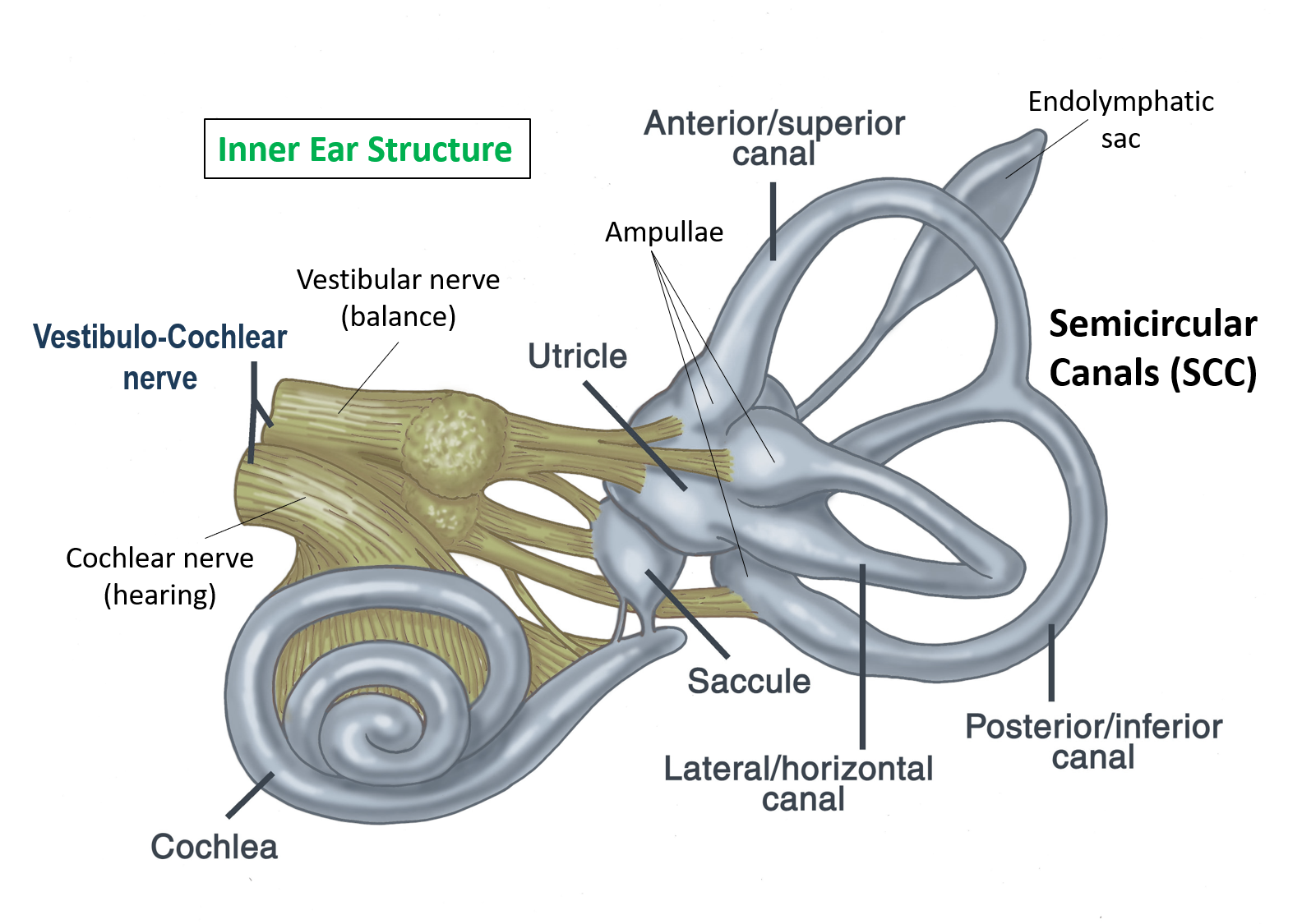


These sense organs of balance in the inner ear sense all kinds of motion and orientation of the body and send those signals via the vestibular nerve to the brainstem where signals from eyes, joints, and muscles are all integrated and checked with learned automatic behaviors in the cerebellum and from memory in the cerebrum of the brain. Any disturbance in this wonderful ‘balancing act’ of so many systems coming together to keep us upright and oriented can cause what is commonly known as vertigo.
Vertigo can be caused by many conditions that broadly can be divided into central and peripheral.
Central vertigo
This results from a condition that affects the brain and its nutrition, structure and function, with disruption of the signaling it gets from peripheral balancing organs. It is usually continuous and affects the overall balance of the patient. Central vertigo most commonly occurs as a result of reduced blood and oxygen supply (ischemia) of the central vestibular structures in the cerebellum, brainstem, or vestibular nuclei. This should be kept in mind especially in the elderly with cardiovascular disease and risk factors. Other causes include multiple sclerosis and migraine. Rare causes include brain trauma, infection and tumors. Vertigo can sometimes be medication-induced especially with a very high dose of anticonvulsants.
Peripheral vertigo
It occurs mainly due to inner ear dysfunction and is much more common and most often the reason for the patient’s visiting doctors.
The doctor may sometimes perform a test to assess peripheral vertigo due to the inner ear (labyrinthine or vestibular causes). This is called the Fukuda-Unterberger Step Test (also called as Unterberger test or Fakuda test) in which the patient is asked to take multiple steps (50-90 steps in 1 minute) in the same place with eyes closed and hands stretched. Deviation to one side of more than 30 degrees suggests the involvement of the labyrinth or vestibule of that side of the inner ear. This test cannot diagnose or rule out central vertigo.
COMMON INNER EAR CONDITIONS CAUSING VERTIGO
BPPV – Benign Paroxysmal Positional Vertigo
As the name implies this is one of the most common conditions causing vertigo in the general population which occurs briefly and periodically in a particular position. That offending position like getting up from the lying position, turning head suddenly side to side, or bending downwards, elicits vertigo along with specific eye movements particular to the semicircular canal affected.
Characteristically, the dizziness and eye movements reduce in intensity when the offending position is repeated multiple times. Currently, BPPV is being diagnosed and treated according to the canal involved
90-95%% of the time it occurs due to displacement of stones in the ear from their natural position, their free-floating inside the problem canal, and settling down at different positions on head movements, thereby causing vertigo. 5-10% of the time, the cause is debris weighing down on the cupula of the semicircular canals, which is supposed to sense angular momentum but due to the debris now senses gravity as well, causing vertigo.
The condition is fairly common and the eye movements give an idea of the offending canal involved. Maneuvers like Dix Hallpike were used to diagnose these conditions. Dix–Hallpike test is performed by quickly lowering the patient to a horizontal, face up position the neck extended 30 degrees. A positive test is indicated by the patient reporting vertigo and the doctor observing the nystagmus (involuntary eye movement). A negative test makes BPPV a less likely diagnosis. The test carries certain risks and contraindications and should only be performed by a trained doctor.
However today, the most accurate diagnosis of BPPV is made by videonystagmography (VNG: video-graphic evaluation of nystagmus eye movements.
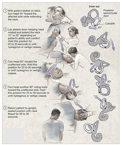
Treatment consists of corrective maneuvers of the head which place the stones floating in the canal back to their original position called canalolith repositioning maneuvers as shown in the image below (like Epley’s Maneuver, Log roll, Barbeque roll, etc). These treat 85-90% of patients along with oral medications that are vestibular relaxants (antihistamines like meclizine and promethazine, antinausea medicines, and benzodiazepine sedatives) as they reduce the intensity of wrong signals being sent to brain.


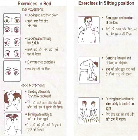
Along with the above, avoiding sudden movements and movements along the axis of the problematic canal, and some vestibular exercises like Cooksey-Cawthorne exercises shown below, are advised to the patient.


Meniere’s Disease
This relatively uncommon disease is caused by increased pressure inside the inner ear system. The inner ear consists of two types of liquids that help transmit sound. Perilymph is the outer liquid that is directly connected to the middle ear. Endolymph is the inner liquid in which the sense organs of balance and hearing and their respective nerves float. The Endolymphatic sac is a small structure that acts as a pressure regulator for endolymph, a collection point for it and helps in the elimination of waste from the endolymph.
Meniere’s disease is caused by problems in the endolymphatic sac which in turn disturbs the homeostasis of the endolymph and its system. This causes increased pressure to be transmitted across the hearing and balance organs that cause a classical triad of symptoms which are –
- fluctuating hearing loss (which is sensorineural – caused by damage to the inner ear or the nerve from the ear to the brain)
- roaring ocean-like tinnitus (noises in the ear)
- vertigo lasting more than 20 minutes
Meniere’s disease is usually of unknown cause and is a disease that has to be managed with both lifestyle changes and medications and at best is to be kept under control. Patients can go on for years before they see another attack of dizziness in Meniere’s if correctly managed.
Patients are generally counseled to avoid driving, piloting planes, or operating heavy machinery, possibly for life. They are asked to keep blood pressure under control and reduce eating salt to keep the endolymph pressure down as most of the symptoms arise because of raised pressure.
Medications (antihistamines like meclizine and promethazine or histamine analog betahistine, antinausea medicines, and benzodiazepine sedatives) are usually given during acute attacks to mitigate that episode. Diuretic drugs may also be prescribed to aid fluid drainage and lower pressure. After the short course of medications is over the patient goes back to the normal routine.
Severe debilitating disease with very frequent attacks of dizziness, falling down, and hearing loss is managed by surgery where the endolymphatic sac is decompressed and shunted for separate drainage.
For diseases not managed by conservative surgery, removal of the inner ear on that side and cutting its nerve supply is done as a final option.
There is a device that gives positive pressure in the ear to relieve episodic attacks of vertigo called the Meniett device shown below. This is a good short-term relief choice.



Meniere’s disease is a long disease with varying severity and multiple management options. Early identification, management and treatment give long-term relief.
Motion sickness
A very common and unpleasant condition occurs when a person is in motion or they perceive they are in motion. This occurs due to a mismatch of sensory inputs given to the brain while in motion. The easiest to identify is car sickness which happens to many people, especially on hilly and twisting roads. Classically patient feels nausea, sometimes vomiting, anxiety, sweating, and dizziness, and this can happen as frequently as every 5 seconds.
Motion sickness can be of three types –
- When motion is felt and not seen – like in car sickness and air sickness.
- When motion is seen and not felt – like in space in a state of constant free fall the person sees the movement but the inner ear balancing system is confused and this causes motion sickness
- When motion is both seen and felt but the integration is wrong – like in centrifugal motion where the motion we see and feel are different
Motion sickness can be troublesome over long journeys and in severe cases can cause fear of travel, dehydration due to vomiting, etc.
A simple treatment for motion sickness is behavioral therapy where the person fixes gaze over the horizon while traveling in a vehicle, limits screen time, gets more fresh air and closing eyes in cases it occurs due to 3D movie screen or virtual reality headsets.
Medication for motion sickness is supposed to be taken 30 min before traveling and soothes the vestibular system so it does not overreact to the motion. Medications available over the counter include oral drugs like cinnarizine, promethazine, and scopolamine (also available as a skin patch).
Vestibular Neuritis and Labyrinthitis
This disease is an inflammation of the inner ear fluid-filled canals (labyrinthitis) and its vestibular nerve (vestibular neuritis). This inflammation can cause severe and continuous dizziness, and sometimes sudden hearing loss in the ear. In severe cases, it causes nausea and vomiting along with vision problems. This inflammation is caused due to infection by viruses like Herpes, the common cold and influenza (flu). In many cases, it can be caused due to autoimmune diseases where the body immune cells attack due to a self-recognition error and cause damage.
Patients typically present with sudden continuous severe vertigo and vomiting with continuous movement of eyes called nystagmus. They are dehydrated and have a history of some recent infection.
Investigations include audiometry which will show some degree of neural hearing loss. Vestibular testing is usually not done because the patient is unable to even stand. In cases of no nystagmus and mild dizziness, Hallpike maneuver is done to elicit nystagmus, after which if no nystagmus is elicited then it points to an infective pathology.
Treatment of this condition consists of emergency intravenous fluids in cases of dehydration with injections to stop vomiting. The patient is given intravenous corticosteroids to reduce inflammation and vestibular corrective and soothing medication like meclizine, dimenhydrinate, betahistine, etc.
The patient is kept on bed rest until the acute episode lasts and then can be mobilized.
Acoustic Neuroma (Vestibular Schwannoma)
This is a rare but benign tumor of the vestibular nerve which carries balance signals from ear to brain or cochlear nerve which carries sound signals from the ear to brain. This is a very slow-growing tumor occurring most commonly from the sheath called the myelin sheath of the nerves. This tumor preferably occurs at the cerebellopontine angle, which is the entry point into the inner ear of the nerves supplying it.
This tumor has a typical gamut of symptoms and 3 or more of these symptoms need to be present to warrant testing for this disease:
- high-pitched ringing sound in the ear (tinnitus)
- frequent vertigo episodes
- slowly progressing hearing loss
- difficulty in understanding speech
- ear fullness
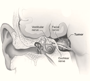


This tumor, although is slowly progressing, can reach large sizes before any symptoms are present. Since the facial nerve is also nearby, which supplies muscles of expression on the face, it can cause weakness of those muscles and facial palsy. Giant tumors can compress the brain and brainstem causing frequent drop attacks (sudden falls without trigger), imbalance issues, headaches, etc.
Testing is done via pure tone audiometry with shows hearing nerve damage. MRI brain with contrast which is the gold standard of diagnosis of this disease helps in surgical planning.
For small tumors with no symptoms wait and watch approach is feasible as the tumor is very slow growing (0.1-0.2 cm per year). Such patients lead their life normally.
For larger tumors with the above-mentioned symptoms, and long-standing tumors, surgery is the treatment of choice which can be done either via the ear itself in case of a deaf ear, or from behind the ear or from the occipital ear. Surgical outcomes are excellent if the tumor is not attached to other nerves coming out of the region.
Patients who are unfit for surgery can undergo stereotactic radiotherapy procedures like gamma knife or cyber knife which use highly focused beams of radiation to remove the tumor.
Bilateral acoustic neuroma occurs in a syndrome called neurofibromatosis 2 which is familial so the family of the patient should also be tested, in such cases.
Dr. Manish Prakash is one of the leading ENT surgeons in Gurgaon. He has trained at the All India Institute of Medical Sciences (AIIMS) after graduation from Maulana Azad Medical College (MAMC), New Delhi. He has also received the DOHNS in Otolaryngology and Head Neck Surgery by the Royal College of Surgeons of England, UK, along with many other awards. Along with general ENT, he has focused his interests in Endoscopic Sinus surgery, STITCHLESS Ear Surgery, Snoring Correction Surgery, PhonoSurgery, and Skull Base surgery. He is a frontrunner for MINIMAL ACCESS ENT SURGERY, and the First ENT Surgeon in Gurgaon to have done Endoscopic Transnasal Repair of CSF Rhinorrhea, Stapedotomy and Endoscopic DCR. He has several research publications and has been a speaker of prominence in medical conferences.
https://www.entgurgaon.com/view-blog.php?title=how-is-a-diagnosis-of-vertigo-is-done
Also read:
Hearing Loss – 5 Important Points of Awareness and Understanding


