

Routine mammography has saved countless lives through early breast cancer detection, but it doesn’t work equally well for everyone. Women with dense breast tissue, those at high risk due to genetic mutations, or patients with inconclusive mammogram results often need more sophisticated imaging approaches. Knowing when to move beyond standard screening—and what options exist—can make the difference between catching cancer early and missing a treatable diagnosis.
The Limitations of Standard Mammography
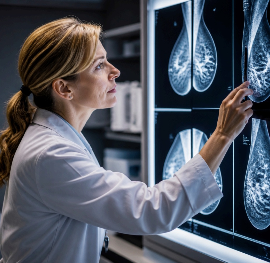
Mammography uses X-rays to detect abnormalities in breast tissue, but dense breast tissue appears white on mammograms—and so do tumours. This overlap reduces sensitivity in approximately 40% of women who have heterogeneously dense or extremely dense breasts. Studies show that mammography detects cancer in about 85% of women with fatty breasts but only 60 to 65% of those with dense tissue.
Dense breasts are not abnormal. Breast density reflects the proportion of fibroglandular tissue relative to fat, and it’s influenced by age, genetics, hormone levels, and body composition.
Younger women typically have denser breasts, which become fattier after menopause. However, density alone increases breast cancer risk by 1.5 to 2 times compared to women with mostly fatty breasts, independent of the masking effect on imaging.
When Breast MRI becomes necessary
Magnetic Resonance imaging (MRI) of the breast uses powerful magnets and radio waves to create detailed cross-sectional images. Unlike mammography, MRI doesn’t rely on tissue density differences. It detects cancer by identifying increased blood flow to tumours after injecting a contrast agent. This makes MRI particularly valuable for high-risk screening and problem-solving in complex cases.
Guidelines recommend annual breast MRI screening for women with a lifetime breast cancer risk of 20% or higher. This includes carriers of BRCA1 or BRCA2 mutations, those with strong family histories suggesting hereditary cancer syndromes, women who received chest radiation between ages 10 and 30, and individuals with certain genetic conditions like Li-Fraumeni syndrome. For these women, MRI detects two to three times more cancers than mammography alone.
MRI also plays a diagnostic role when mammography and ultrasound produce uncertain findings. A palpable lump that doesn’t show on conventional imaging, bloody nipple discharge without an obvious cause, or a newly diagnosed cancer requiring surgical planning all warrant
MRI technology excels at determining tumour size, detecting additional disease in the same breast, and screening the opposite breast for occult cancer.
The Challenge of MRI-Detected Abnormalities
Greater sensitivity comes with trade-offs. Breast MRI has a false-positive rate of 10 to 15% in screening populations, meaning it identifies suspicious areas that turn out to be benign. When MRI reveals a concerning finding that wasn’t visible on mammography or ultrasound, confirming whether it represents cancer requires tissue sampling under MRI guidance.
This is where advanced biopsy techniques become essential. Cleveland Clinic Abu Dhabi’s Breast Health & Cancer Program offers comprehensive diagnostic services including MRI guided breast biopsy for patients in Abu Dhabi who need tissue diagnosis of abnormalities visible only on MRI.
The procedure uses real-time MRI imaging to guide a needle to the precise location of the suspicious area, allowing pathologists to determine whether cells are cancerous, precancerous, or benign. This capability closes a critical diagnostic gap for women whose cancers would otherwise go undetected or require invasive surgical biopsy.
Navigating Dense Breast Notifications
Many regions now require radiologists to inform women when mammography reveals dense breast tissue. These notifications often create confusion and anxiety. Having dense breasts doesn’t mean you have cancer or even that cancer is likely, but it does warrant a conversation with your doctor about whether supplemental screening makes sense for your individual situation.
Not every woman with dense breasts needs MRI. The decision depends on additional risk factors: family history, personal history of breast biopsies showing atypical cells, age at first menstruation and first childbirth, and genetic testing results.
Ultrasound serves as a middle ground for some women—it’s less expensive and more widely available than MRI, though it detects fewer cancers and has variable accuracy depending on the operator’s skill.
Women should understand that adding supplemental screening increases both cancer detection and false alarms.
Breast care awareness and education help patients make informed choices about screening intensity based on their personal values around early detection versus potential anxiety from additional testing.
What to Expect from Breast MRI Screening
A breast MRI exam typically takes 30 to 45 minutes. You lie face down on a padded table with openings for your breasts, which hang into a dedicated coil that captures the images. An intravenous line delivers gadolinium contrast midway through the scan. The machine produces loud thumping and buzzing sounds, so you’ll wear earplugs or headphones.
Timing matters for premenopausal women. MRI should occur during days 7 to 14 of the menstrual cycle when hormone levels minimize background breast tissue enhancement. This reduces false positives caused by normal hormonal changes. Postmenopausal women not taking hormone therapy can have MRI at any time.
The main contraindications are severe kidney disease, which increases risks from gadolinium contrast, and certain metallic implants incompatible with strong magnetic fields. Most modern pacemakers, defibrillators, and surgical clips are now MRI-safe, but confirmation is essential before scheduling.
Integrating MRI into Your Screening Strategy
High-risk women benefit most from alternating MRI and mammography at six-month intervals, providing imaging every six months rather than annually. This approach balances the strengths of both technologies: mammography’s ability to detect calcifications that MRI might miss, and MRI’s superior soft tissue contrast and sensitivity in dense breasts.
Cost and access present real barriers. Breast MRI costs several times more than mammography, and insurance coverage varies. High-risk screening MRI is generally covered for women meeting established criteria, but supplemental screening for density alone often isn’t. Facilities offering breast MRI are less common than mammography centres, particularly in rural areas, which can mean travelling significant distances for annual screening.
Despite these challenges, outcomes justify advanced imaging for appropriate patients. Cancers detected through high-risk MRI screening are typically smaller and less likely to have spread to lymph nodes compared to those found through symptoms. Earlier detection translates to less intensive treatment options and better long-term survival.
Moving Forward with Confidence
Understanding your breast cancer risk profile enables better screening decisions.
- If you have a strong family history of breast or ovarian cancer, ask your doctor about genetic counselling and risk assessment.
- If mammography reports dense tissue, discuss whether your overall risk level warrants additional imaging.
- If MRI reveals an abnormality requiring biopsy, know that MRI-guided tissue sampling can provide definitive answers.
Breast imaging technology continues advancing, with artificial intelligence algorithms improving accuracy, abbreviated MRI protocols reducing scan times and costs, and contrast-enhanced mammography offering a middle option between conventional mammography and full MRI.
The goal remains unchanged: detecting cancer when it’s most treatable while minimizing unnecessary anxiety and intervention. Matching the right imaging approach to each woman’s individual circumstances makes that goal achievable.
Also read