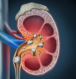
Urinary stones (renal calculi) can form anywhere in the urinary tract, and cause obstruction leading to pain and difficulty in urination. They also increase the risk of urinary tract infections. Long-standing or recurrent stones can compromise renal function.
WHO IS AT RISK?
- Dehydration from not drinking enough fluid
- A diet too high in protein, oxalates, salt and sugar
- Being overweight or obese
- Hormonal diseases that cause increased calcium loss in urine
- Inflammatory bowel disease (IBD) or after gastric bypass surgery

TYPES OF URINARY STONES
There are 5 main types of urinary stones (renal calculi):
Calcium Oxalate Stones
They are the most common type of kidney stones and form when the urine contains low levels of citrate and high levels of calcium along with oxalate or uric acid. Calcium oxalate stones are linked with foods high in oxalate, which naturally occurs in foods like beets, black tea, chocolate, nuts, potatoes, and spinach. IBD and gastric bypass surgeries increase the risk of oxalate stones.
Calcium Phosphate Stones
Calcium phosphate kidney stones are less common and often co-exist with calcium oxalate stones due to increased calcium in urine. They may also be formed in a condition called renal tubular acidosis.
Struvite Stones
More common in women, struvite stones form as a result of certain types of urinary tract infections with bacteria that produce ammonia that makes the urine alkaline. These stones are made of magnesium, ammonium, phosphate and calcium carbonate. They tend to grow quickly and become large, sometimes occupying the entire kidney. Left untreated, they can cause frequent and sometimes severe urinary tract infections and loss of kidney function.
Uric Acid Stones
More common in men, uric acid stones tend to occur in people who don’t drink enough water or have a diet high in animal protein, which makes their urine more acidic. They are also more common in people who have gout, a family history of kidney stones, or in those who have had chemotherapy.
Cystine Stones
Cystine stones are caused by a hereditary genetic disorder called cystinuria that can lead to excessive amounts of the amino acid cystine collecting in the urine.
SYMPTOMS
Stones go undiagnosed unless symptoms like severe pain (renal colic) due to obstruction occur. Depending on the location of the stone, the pain can be in the lower back or groin.
Urination becomes difficult and painful, and there may be increased frequency and urgency of urination.
There may be accompanying nausea and vomiting due to the pain.
Sometimes blood may be present in urine (hematuria).
Urinary tract infections are frequent, causing fever, discomfort, and burning while urinating.
DIAGNOSIS
After a complete history and clinical examination to detect site of pain, renal function tests are ordered that include both blood and urine tests.
A simple X-ray, called a KUB (Kidney Ureter Bladder) may reveal the stone, or else a CT scan or an intravenous pyelogram (IVP) may be performed (by injecting a contrast dye).
TREATMENT
Increase fluid intake
The most important and basic thing to prevent stone formation is to drink and urinate more than two liters per day. Drink a glass of water every hour consciously.
Dietary modification
Salt intake
When excess sodium is excreted in the urine, calcium is also excreted proportionally. So, reduce salt intake to less than 2 grams of sodium per day.
Calcium and Oxalate
There is a misconception that one must restrict their calcium intake if they have calcium stones. This should not be done and calcium with vitamin D should be consumed in the required normal levels to maintain healthy bones and body function. Foods rich in oxalate may be restricted.
Normally calcium and oxalate bind together in the intestine and are eliminated from the body. If calcium is low in diet, the oxalate will be reabsorbed by the body and eliminated through the urine which increases the risk of calcium oxalate stones.
Proteins
Reduce animal proteins especially red meats in diet.
Citrus juices
Citrate is a molecule that binds to calcium in the urine, preventing calcium from binding to oxalate or phosphate and forming a stone. One can increase citrus fruit juices in diet.
Medicines
Pain medicines
These are given to control the pain till other measures come into action.
Diuretic medicines
These drugs help to decrease urine calcium excretion (thiazide diuretics). They also help to keep calcium in the bones, reducing the risk for osteoporosis. The most common side effect of thiazide diuretics is potassium loss, so the doctor will prescribe a potassium supplement to go along with the thiazide diuretic.
Citrate supplementation
The doctor may evaluate electrolyte levels of sodium and potassium in blood and prescribe potassium citrate or sodium citrate supplements accordingly.
Anti Gout Medicines
These may be given in case of patients who have increased uric acid in the blood, and gout. These drugs include allopurinol and febuxostat.
Antibiotics
Urinary tract infections should be treated with appropriate antibiotics after urine test, microscopy and culture.
Procedures
There are some instances when a kidney stone is left untreated, as in small ones (less than 5 mm) and not causing any pain. These usually pass on their own after falling into the ureter. Such stones may be observed with periodic X-rays to watch for growth or changes.
The following stones should be treated with interventions:
- Stone causing pain not controlled with oral pain medication
- Presence of severe nausea and vomiting
- Stones causing recurrent or severe urinary tract infections
- Stones blocking urine flow
- Stones larger in size than 5 mm are unlikely to pass on their own
- Staghorn stone – Extremely large stones that grow to fill the inside of the kidney, and can cause kidney failure.
- Stones not responding to dietary, or medical management.
- Occupational requirements for certain professions
ESWL (Extracorporeal Shock Wave Lithotripsy) is a non-invasive procedure that breaks down stones in parts of the urinary system, by using shock waves aimed at stones, with the help of X-ray or ultrasound. Small broken-down pieces of stones in the kidneys and ureter then pass on their own in urine.
Ureteroscopy usually done in a clinic under general anesthesia, involves the passage of a small flexible telescope, called a ureteroscope, through the urethra and bladder and up the ureter to the point where the stone is located. Small stones are removed with a basket-like device which large stones are lasered and the pieces are then removed. A stent may be left in the ureter for about a week to prevent blockage due to post-procedure swelling or inflammation.
PERC (Percutaneous) nephrolithotomy is a procedure done in hospitals under general anesthesia. It creates a passageway from the skin on the back to the kidney. A surgeon uses special instruments passed through a tiny tube in the back to locate and remove stones from the kidney. Percutaneous nephrolithotomy is used most often for larger stones (>2cm) and staghorn stones or when less-invasive procedures are not feasible.
It is important to be aware and discuss the risks of surgical interventions with your treating doctors like bleeding, swelling, infection, injuries, and post recovery urinary difficulties, stone piece remnants, and general convalescence.
Also read:
Urinary Tract Infection (UTI) – urethritis, cystitis, ureteritis, pyelonephritis
Chronic Kidney Disease (CKD) – 5 Key Points of Understanding

